All published articles of this journal are available on ScienceDirect.

Surge Capacity Treatment for Open Fractures in the Gaza Strip: Case Reports
 iD
Shahd Abu Hamad2
Khulood Abul Qaraya2
Theresa Farhat2
Ghassan Abu-Sittah2
Mahmoud Matar3
Sadiq Abdelall4
Anthony M. J. Bull1
iD
Shahd Abu Hamad2
Khulood Abul Qaraya2
Theresa Farhat2
Ghassan Abu-Sittah2
Mahmoud Matar3
Sadiq Abdelall4
Anthony M. J. Bull1
Authors Info & Affiliations
Abstract
Introduction/Background
Treating open fractures in conflict zones, such as the Gaza Strip, is severely influenced by supply chain disruptions and shortages of medical devices. This study addresses this by evaluating the clinical application of a low-cost, locally manufacturable external fixator developed by Imperial College London. It contributes to the literature by demonstrating the viability of frugal innovation in providing surge capacity for complex trauma in an active war zone with severe resource constraints.
Case Presentation
Two male patients, aged 21 and 13, presented to Shifa Hospital in Gaza with Gustilo-Anderson Type 3B and Type 1 open tibial fractures, respectively. Within 24 hours of admission, both underwent wound debridement and definitive stabilization using the Imperial External Fixator (iFix). Clinical follow-up assessed fracture union and device stability. Both patients achieved successful soft tissue healing and complete bone union (at 7 months and 3 months, respectively), without device failure or loosening. One patient experienced a pin-site infection, which resolved with antibiotics. Surgeons reported that the device provided satisfactory stability and intraoperative modularity comparable to commercial standards.
Conclusion
The low-cost Imperial external fixator proved effective as a definitive treatment for open tibial fractures in an austere, conflict-affected environment. The successful outcomes underscore that locally manufacturable, cost-effective medical devices can reliably bridge critical supply gaps, offering a scalable solution for orthopaedic surge capacity in humanitarian crises.
1. INTRODUCTION
In March 2024, the British Red Cross published an article titled “Broken healthcare and disease in Gaza could kill more than conflict” [1]. This is not a new phenomenon; crippled healthcare systems and shortages of medical supplies are known patterns that have been observed repeatedly in many conflict zones [2, 3]. The authors have witnessed and/or experienced this issue first-hand in countries such as Syria, Ukraine, Iraq, and Palestine (Gaza Strip). Over the past several decades of conflict in the Gaza Strip, there have been tens of thousands of casualties, many of whom had open fractures. For instance, during the Great March of Return (GMR) demonstrations between March 2018 and October 2020, there were approximately 320 fatalities and over 33,000 injuries reported. Among these, 41% sustained Gunshot Wounds (GSWs) to the lower limb, primarily open tibial fractures [4, 5]. Managing these injuries is challenging, especially in conflict zones like the Gaza Strip. Limited resources and infrastructure strain healthcare systems, hindering comprehensive treatment, despite the need for multiple surgeries and prolonged recovery periods.
A recent study involving 244 individuals afflicted with open tibial fractures resulting from GSWs, predominantly young adult males in the Gaza Strip, revealed that nearly half had Gustilo-Anderson type 3B fractures. Furthermore, over two-thirds of cases had > 10mm of bone loss [4]. Most patients required multiple surgeries, including rotational flaps and bone grafts, with around 41.2% needing dual fixators during follow-up. Non-union occurred in 53% of cases, underscoring the urgent need for specialized trauma care resources to address the significant burden of lower limb injuries from violence in the Gaza Strip [4]. The ongoing war (started in October 2023) has surpassed all previous conflicts in the Gaza Strip in terms of casualties, many of whom have been children, where poorly treated fractures could lead to life-lasting complications worse than in adults [6]. Between 7 October 2023 and 1 May 2024, over 77,765 Palestinians have been injured in the Gaza Strip, and fractures are among the main types of injuries sustained due to heavy bombing [7, 8]. The use of an external fixator as a first-line treatment for open long-bone fractures is well established, particularly in austere environments and in Low- and Middle-Income Countries (LMICs) [9-11]. This is a temporary solution, and depending on the wound and the patient's condition, a definitive internal treatment is implemented after a few weeks, enhancing the patient's mobilization and the rate of bone union. However, various factors, including the risk of infection, the availability of internal fixation techniques, the patient's general condition, and other socio-economic factors, affect the conversion to internal fixation, leading to the use of external fixation as a definitive treatment in many low-income settings. In those austere settings, low-quality devices are often reused multiple times due to the high cost of commercial external fixators [11]. In regions such as the Gaza Strip, where importing goods is severely restricted, the situation is even worse, and the shortage of medical supplies is a critical problem [2, 3, 11]. On different occasions, such as the ongoing war, importing medical supplies, even through humanitarian channels, is extremely difficult/slow, if possible at all. Therefore, a locally manufactured solution with surge capacity (i.e., addressing a sudden need for large numbers of devices) could be a game-changer.
To address this problem, Imperial College London (ICL) developed a low-cost external fixator, iFix, and a toolkit to manufacture it locally in an austere environment [2]. The fixator has undergone testing to meet international standards (ASTM F1541-17 – 17 Standard Specification and Test Methods for External Skeletal Fixation Devices). As detailed in Saeidi et al., despite similar performance, iFix costs a fraction of the cost of commercial external fixators due to its simple manufacturing technique and the materials it is made of [2]. Additionally, a clinical trial was conducted at tertiary care hospitals in Sri Lanka involving 17 patients across various configurations to evaluate the device's initial clinical performance and assess outcome measures. This trial revealed the device’s excellent stability and minimal complications, and received positive feedback from surgeons and patients [11].
This study aimed to evaluate the efficacy of this external fixator in a conflict setting, the Gaza Strip, for treating open fractures in the tibia as a primary and definitive treatment. This case study was performed at the Shifa Hospital.
2. CASE PRESENTATION
2.1. Study Design
This case study involved the enrolment of two patients with open tibial fractures who underwent external fixation with the Imperial external fixator at the Shifa Hospital in the Gaza Strip, with subsequent assessment of their outcomes during follow-ups. Only two eligible cases were included, as this report reflects the initial phase of the study using UK-manufactured investigational devices before the planned expansion with locally manufactured devices in Gaza was interrupted by war-related disruptions.
The study was initially designed in two phases: the first using devices made in the United Kingdom (UK), and the second phase using devices made in Gaza. In the cases studied, the devices were manufactured in London using Computer Numerical Control (CNC) machining. Technical information about the device used in this study is available in a previously published journal article [2]. The device used in this study is investigational and has not yet been approved. After manufacturing, the devices were shipped to the Gaza Strip through Medical Aid for Palestinians (MAP), which took 10 months merely due to the extremely slow approval process by the Israeli government for all the goods imported to the Gaza Strip, including the humanitarian supplies sent by humanitarian organisations. This resulted in a significant delay in the study's start.
Even though the use of the device is similar to that of commercial external fixators, to ensure correct implementation and as part of capacity building, two training workshops were held by the senior surgeon (MM) at Shifa Hospital, attended by 21 residents and consultants.
2.2. Studied Cases
The study included two patients, aged 21 and 13, both with compound tibial fractures, meeting the inclusion criteria of being admitted to the hospital within a week after injury, not requiring joint-spanning external fixation, being able to provide consent (informed consent was obtained in writing), and not having a history of previous tibial injury. For the second patient, aged 13, written informed consent was obtained from the patient's parent/legal guardian. These cases necessitated the application of external fixators based on surgical assessments, followed by the utilization of the ICL low-cost external fixator.
2.3. Outcome Measures
The study examined complications associated with the ICL low-cost external fixator and evaluated fracture stability and healing at 2, 4, 8, and 12 weeks following application, and then performed monthly assessments until the device was removed. Outcomes were assessed clinically and radiographically during follow-up and included pin-tract infection, pin loosening, loss of fixation, construct stability, soft tissue healing, and fracture union; formal patient-reported functional outcome scores were not collected in these two cases.
2.4. Data Collection and Variables
Data collection began upon application of the external fixator and continued for 12 weeks, or until the device was removed. It included demographics, injury details (mechanism of injury, severity scores, fracture patterns), follow-up assessments (pin-site infections, loosening, soft-tissue healing, radiological findings), and surgeon feedback on setup, pin placement, configuration, conformity, stabilization, confidence, injury risk, and overall impression. Clinical, radiological, and fixator-specific data (i.e., slippage and clamp deformation) were gathered at each follow-up.
3. RESULTS
Patient 1, a 21-year-old male smoker, was admitted to Shifa Hospital within 3 hours of sustaining an injury in a road traffic accident. He presented with a complex lower limb trauma, including an open tibial fracture classified as Gustilo-Anderson Type 3B. The injury involved a left intersegmental tibia fracture, serial metatarsal and navicular fractures, and left foot compartment syndrome. Despite minimal bone loss (< 1cm), the injury required immediate intervention. Within less than 6 hours of admission, wound debridement and unilateral uniplanar external fixation were performed by an orthopaedic surgeon (Fig. 1a), with secondary soft tissue closure. A post-operative photo of the limb is shown in Fig. (1b). No weight-bearing was allowed initially to promote healing. Intravenous (IV) antibiotic therapy included cefazolin and gentamicin. At the 4-week follow-up, no complications were observed. Radiological examination revealed clear evidence of fracture alignment and soft tissue healing following skin graft. However, there was still no indication of callus formation or union at this stage. The device was ultimately removed after 7 months once the full bone union was achieved.
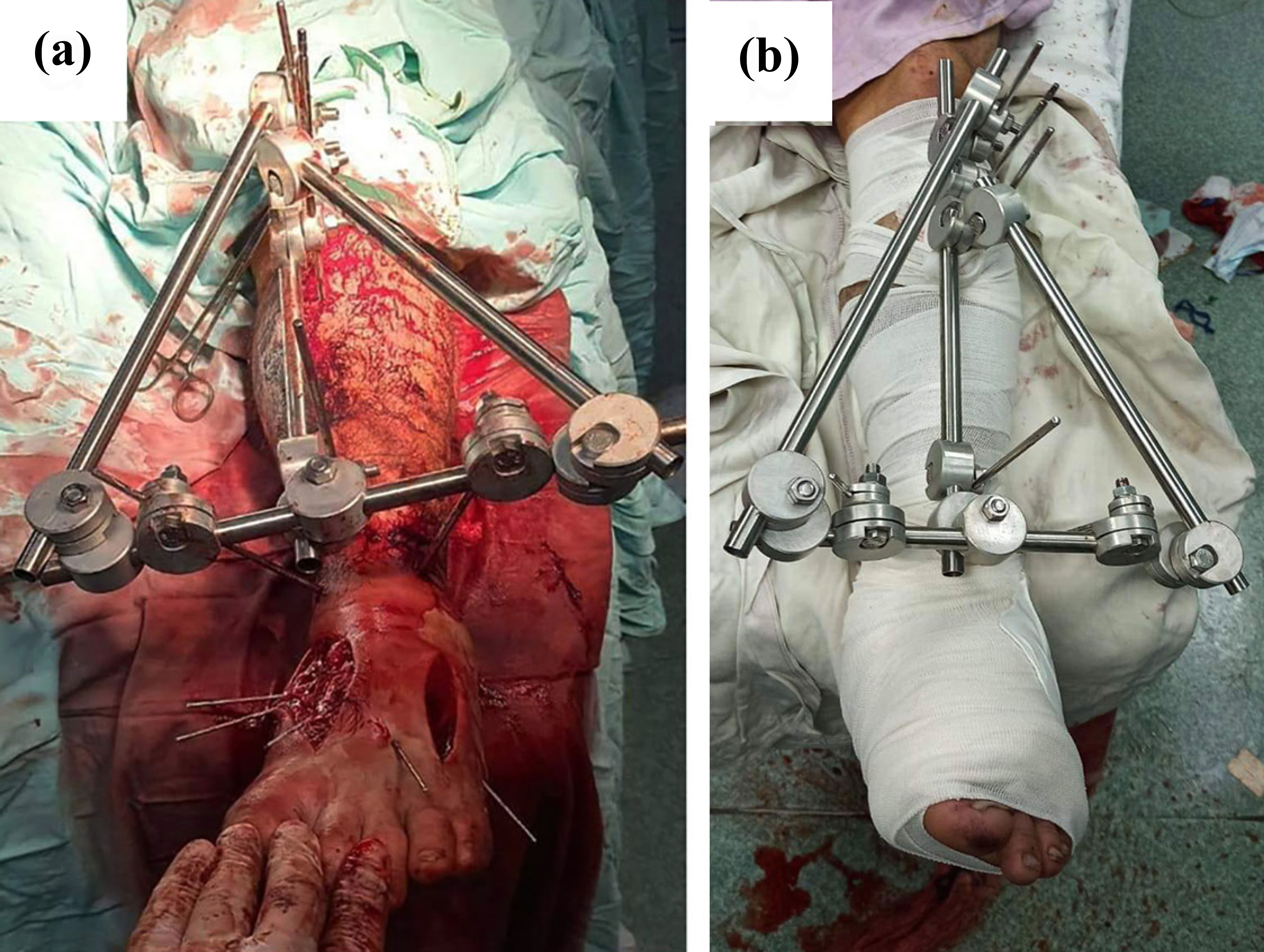
a) Intraoperative, and b) Post-operative photos of external fixation of the tibia.
Patient 2, a 13-year-old male, presented to the emergency room within 2 hours of sustaining an injury while playing football. He was diagnosed with a Gustilo-Anderson Type 1 open tibial fracture characterized by a fragmentary pattern and no bone loss (Fig. 2a). Within 2 hours of admission, wound debridement and external fixation with a unilateral uniplanar configuration were conducted by an orthopaedic surgeon (Fig. 2b).
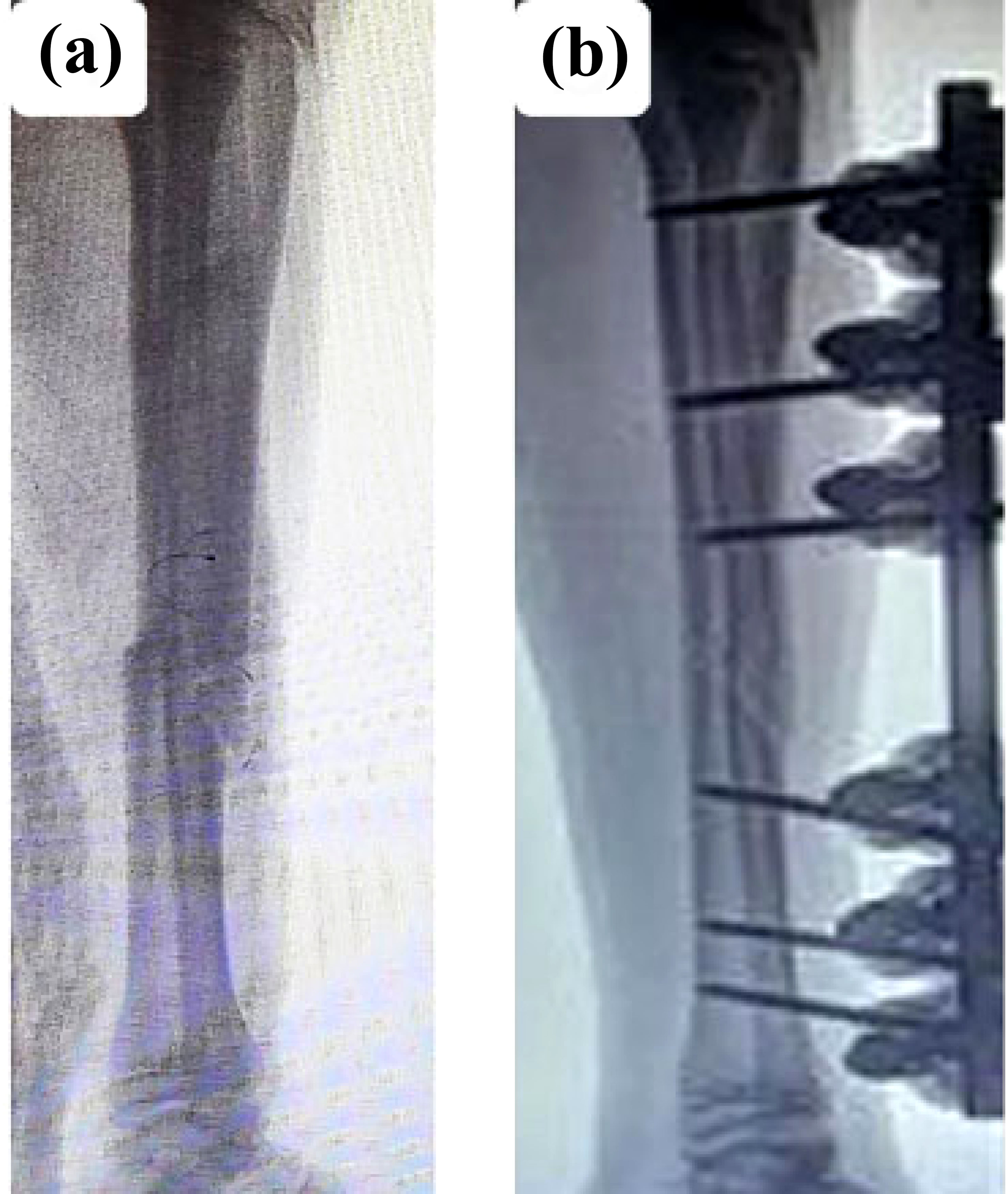
a) Pre-operative, and b) Post-operative X-rays of the tibia.
No weight-bearing was allowed initially to promote healing. Primary soft tissue closure was achieved, albeit with noted skin bullae. Intravenous antibiotic therapy, Zinacef, was administered. Following the two-week follow-up, no immediate complications were noted; however, a pin-site infection, Checketts grade 2, developed during the subsequent evaluation. This infection was treated with regular dressing changes and a regimen of oral antibiotics, including trimethoprim-sulfamethoxazole and fusidic acid ointment. Despite visible callus formation, bone union had not yet been achieved at the time of assessment. After one month from the initial injury, the ongoing pin-site infection required a repeat course of antibiotics. This regimen was continued for 1.5 months. After approximately 3 months, the device was removed when a complete bony union was observed (Fig. 3a and b), and plaster of Paris was applied. This was also removed after 2 weeks.
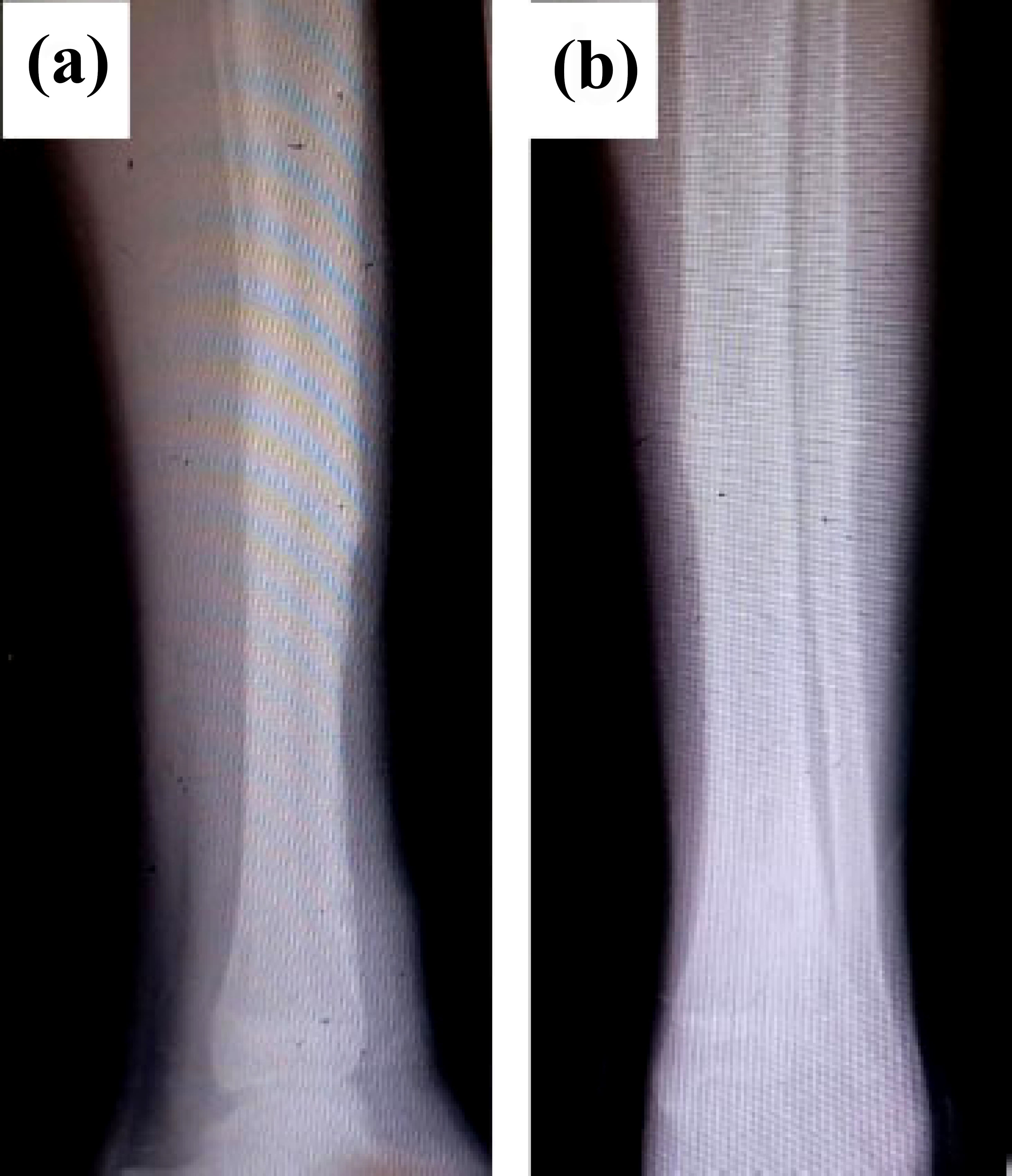
a) Lateral, and b) Antero-posterior view of the fracture after fixator removal.
In both cases, the external fixator provided a stable construct, aiding in soft-tissue management and fracture alignment. Notably, neither case exhibited instances of pin loosening or slippage at the connecting rod to the large split clamp joint. Additionally, the construct's stability was evident, as it could be lifted independently by rods. Surgeons' feedback revealed satisfactory stabilization with the external fixator in both cases. Despite longer setup times than those of alternative methods, the fixator demonstrated excellent fracture stabilization capability. Additionally, it provided satisfactory freedom of pin placement and facilitated the setup of modular configurations, with the conformity of clamps, rods, and Schanz screws ensuring precise stabilization. Surgeons acknowledged the moderate risks associated with handling components but expressed satisfaction with the external fixator's overall performance.
4. DISCUSSION
Based on many decades of conflict in the Gaza Strip, where a new war can break out at any moment, a cost-effective treatment with the purpose of primary definitive fixations is highly needed. However, given the unique circumstances of the Gaza Strip, where the Israeli government could even cut water, fuel, and electricity, as has been done in the ongoing war, importing life-saving medical equipment is not a suitable option. Therefore, local manufacturing of medical devices would be the only reliable option. As a comparator, the authors observed the crucial role of local manufacturing capacity, which was distinctive in supplying medical devices, including external fixators, in Türkiye compared to Syria during the recent earthquake. Local partners in Türkiye informed the authors that thousands of external fixators were manufactured in the first week of the earthquake, while a surgeon based in northwest Syria told the authors that all their devices were used in the first 48 hours, and hundreds of patients were left untreated without access to external fixators across the region.
The present study evaluated the efficacy of a locally manufacturable, low-cost external fixator for the treatment of open tibial fractures in conflict settings. The key findings demonstrated successful fracture stabilization, soft tissue healing, and bone union in two patients. No instances of pin loosening, slippage, or loss of fixation were observed, highlighting the construct's stability. Surgeons reported satisfactory stabilization, freedom of pin placement, and the ability to establish precise modular configurations. The positive outcomes observed in this study align with the well-established role of external fixation as a viable treatment option for open fractures, particularly in austere environments and low-resource settings [12, 13]. The absence of pin loosening or slippage at the connecting rod-clamp interface underscores the construct's stability and aligns with the principles of fracture stabilization outlined in the literature [12]. Furthermore, the surgeons' feedback regarding satisfactory stabilization, freedom of pin placement, and modular configurability reinforces the external fixator's versatility and adaptability, as highlighted by koussai et al [14]. The successful fracture healing observed in this study aligns with the role of the blood supply in the healing process, highlighted by Kumar and Narayan [15].
The setup times reported by surgeons in this study are consistent with the technical challenges associated with external fixator applications in austere environments, as acknowledged by Clasper and Phillips [16]. However, the authors emphasize that the benefits of such frugal technology with surge capacity outweigh the potential drawbacks of longer setup times. The use of a locally manufacturable, low-cost device in this study further highlights the importance of frugal innovation and resource-efficient healthcare solutions for resource-constrained settings, as observed in the previous trial of this device in Sri Lanka and the deployment of this device in Ukraine at the beginning of the ongoing war [2, 11]. In Sri Lanka, the device was utilized in tertiary care hospitals, demonstrating excellent stability and minimal complications, and receiving positive feedback from both surgeons and patients. It proved effective in orthopaedic trauma care and was successfully sterilized and reused. Despite limitations, such as the lack of a comparator device, the trial highlights its potential for deployment in civilian and humanitarian settings, bolstered by its open-source design and published test results. Overall, the findings of this study are consistent with the existing body of knowledge on the efficacy and principles of external fixation in managing open fractures, particularly in austere environments. The technical details and recommendations provided in the cited studies offer valuable insights and reinforce the importance of adhering to established surgical techniques and biomechanical principles for successful outcomes.
5. LIMITATIONS
While this case study's findings are promising, it is important to acknowledge the limitations. Firstly, the small sample size of two patients limits the generalizability of the results. A larger-scale study with the locally manufactured devices at the Islamic University of Gaza was cancelled due to the war. It is worth noting that the device was successfully manufactured in the Gaza Strip using the Imperial toolkit, as shown in Fig. (4).
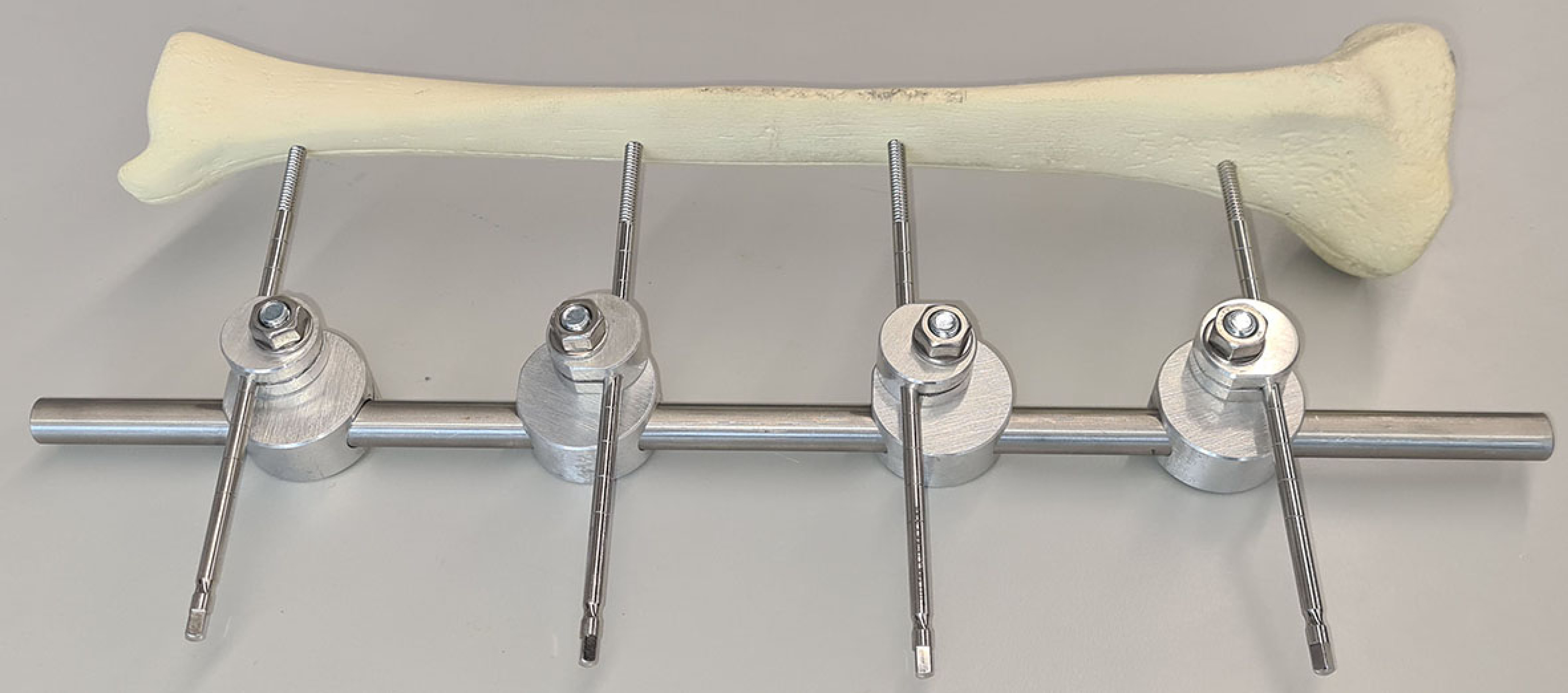
Photo of the locally made external fixator in Gaza, using the Imperial toolkit.
Secondly, the study did not compare the outcomes with alternative treatment methods, such as intramedullary nailing. This is because the nailing technique is unavailable in the Gaza Strip for multiple reasons, including limited availability, infection risk, and a lack of required expertize and fluoroscopy equipment. Thirdly, the follow-up duration was limited to the time required for bone union, precluding the assessment of long-term outcomes and potential complications. Longer-term studies are warranted to evaluate the external fixator's performance regarding functional recovery, quality of life, and potential late-onset complications. Finally, the study was conducted in a specific austere environment, which may introduce potential biases related to resource availability, surgical expertise, and patient characteristics. Replicating the study in different settings would enhance the generalizability of the findings.
6. IMPLICATIONS AND FUTURE DIRECTIONS
The successful implementation of a frugal external fixator in a conflict setting has significant implications for improving access to essential trauma care in resource-constrained environments. This study demonstrates the feasibility of developing and deploying such medical devices to address critical healthcare needs, particularly in regions where importing medical supplies is challenging due to logistical or geopolitical constraints. Future research should focus on expanding the scope of the study to include a larger patient cohort, diverse injury patterns, and comparative analyses with other treatment methods. Long-term follow-up studies are necessary to evaluate the external fixator's performance in terms of functional outcomes, quality of life, and potential late-onset complications. Further research is warranted to explore the local manufacturing of other essential medical devices and surgical instruments, leveraging the principles of frugal innovation and resource-efficient healthcare solutions. Such efforts could help build resilient, self-sufficient healthcare systems in low-resource settings, ultimately improving access to quality care for underserved populations.
CONCLUSION
This study highlights the effective use of external fixation in the management of open tibial fractures in a low-resource setting. Despite the limitations in patient demographics and injury characteristics, timely intervention and meticulous wound care facilitated favourable clinical outcomes. Surgeon feedback underscores the importance of further optimizing external fixation protocols to enhance efficiency without compromising patient care.
AUTHORS’ CONTRIBUTIONS
The authors confirm contribution to the paper as follows: M.S.: Supplying the devices and write-up; S.A.H.: Data collection and write-up; K.A.Q.: Data collection and write-up; T.F.: Ethical approval, write-up, and coordination; G.A.S.: Coordination, clinical protocol, and review; M.M.: Surgical procedures and review; S.A.: Local manufacturing and review; A.M.J.B.: Lead investigator and review.
LIST OF ABBREVIATIONS
| CNC | = Computer Numerical Control |
| GMR | = Great March of Return |
| GSWs | = Gunshot Wounds |
| iFix | = Imperial External Fixator |
| ICL | = Imperial College London |
| IV | = Intravenous |
| LMICs | = Low- and Middle-Income Countries |
| MAP | = Medical Aid for Palestinians |
| UK | = United Kingdom |
ETHICS APPROVAL AND CONSENT TO PARTICIPATE
The study involved human subjects and received ethical approval from the Helsinki Committee for Research Ethics in the Palestinian Ministry of Health in Gaza and the health authorities. Approval/reference number: PHRC/HC/921/21.
HUMAN AND ANIMAL RIGHTS
All human research procedures followed were in accordance with the ethical standards of the committee responsible for human experimentation (institutional and national), and with the Helsinki Declaration of 1975, as revised in 2013.
CONSENT FOR PUBLICATION
Written informed consent was obtained from the first patient. For the second patient, who was 13 years old, written informed consent was obtained from the patient’s parent/legal guardian.
AVAILABILITY OF DATA AND MATERIALS
The data supporting the findings of this article are available within the article.
FUNDING
This research was funded by the NIHR (16/137/45 and NIHR133314) using UK international development funding from the UK Government to support global health research. The views expressed in this publication are those of the author(s) and not necessarily those of the NIHR or the UK Government. This research was also funded by the Imperial College London, United Kingdom EPSRC Impact Acceleration Account EP/R511547/1.
ACKNOWLEDGEMENTS
Declared none.