All published articles of this journal are available on ScienceDirect.
Engineering Approach to Identifying Patients with Colon Tumors on the Basis of Electrophotonic Imaging Technique Data
Abstract
Background:
Colonic neoplasms are quite a serious problem today. Screening methods play an important role in diagnosing the disease. Colorectal cancer screening is a complex undertaking, having various options, which require a lot of efforts both from the doctor and from the patient, including the use of sedatives and the necessity of the presence of an assistant for some procedures such as colonoscopy. This is why it is very important to find a method by which one can make a diagnosis quickly, easily, and painlessly.
Methods:
The ability to identify patients with tumors of the colon using the Electrophotonic Imaging (EPI) technique, as well as using it for differential diagnosis of tumors of the colon by their morphology, size and quantity was investigated. Selection of the most significant parameters of the EPI-graphy for the separation of the control group and the group of patients with tumors of the colon was developed. 137 people were studied with the EPI camera, with ages ranging from 16 to 86 years, including 49 males and 88 females. Based on the results of the colonoscopy and histological findings all subjects were divided into 2 groups: control group of 55 people, 9 males, 46 females; and patients with tumors (benign or malignant) of the colon - 82 people; 40 males and 42 females. Then all subjects were divided into smaller groups based on morphology, size, number of tumors and localization.
Results:
Based on the identified indicators decision rules to determine the patients with tumors of the colon were constructed. The specificity of the resulting function was 80.0% and sensitivity 75.6%. Decision rule was built as well with logistic regression. The specificity of the resulting function was 78.2% and sensitivity 90.0%. The accuracy of this approach was higher than using discriminant analysis.
Conclusions:
The results of this study have proven the ability to identify patients with tumors of the colon using EPI technology, as well as use it for differential diagnosis of tumors of the colon by their morphology, size and quantity. EPI testing is non-invasive, takes less than five minutes, and equipment is relatively cheap and accessible in mass production. This opens up good prospects for further research for implementation as a first step of the screening process. This paper presents the pilot study developing methodological approach to the GDV data processing. That is why we tried different methods of data processing. At the same time we do not pretend to develop a diagnostic method – sample size is too small for this, and other cancer types were not studied. Further research is needed.
1. INTRODUCTION
According to the World Health Organization, each year in the world more than 500 thousand cases of colorectal cancer (CRC) are registered. The highest incidence observed is in the United States, Canada, Western Europe and Russia. Only 25% of the malignant tumors is registered at the first stage; the percentage of patients dying from CRC, as a percentage of deaths from all types of cancer in 2013, was 11.3% for men and 15.9% for women [1]. To date, in Russia, colorectal cancer occupies a leading position. Over the past 20 years, in the Russian Federation, colon cancer has moved from 6th to 3rd highest level of incidence of all forms of the disease [1].
Colonic neoplasms are quite a serious problem today. Screening methods play an important role in diagnosing the disease. There are many ways to determine colorectal cancer such as the gemokult test, immunoassay, colonoscopy, computed tomography (CT), magnetic resonance imaging (MRI), and positron emission tomography (PET) (http://www.globality-health.com/en/). Colorectal cancer screening is a complex undertaking, having various options which require additional effort from the patients (collection of samples for the determination of fecal occult blood, preparation for colonoscopy, etc.), including the use of sedatives and the necessity of the presence of an assistant for some procedures such as colonoscopy [2]. This is why it is very important to find a method by which one can make a diagnosis quickly, easily, and painlessly. One method which can be used to accomplish this is Electrophotonic Imaging (EPI) based on gas discharge visualization (GDV) [3].
The purpose of this research was to identify statistically significant evidence of the EPI technology’s ability [3] to detect individuals with tumors of the colon, as well as to assess the possibility of differential diagnosis of colon tumors with the assistance of EPI. To achieve these goals, the following tasks were determined:
1. Select the most significant parameters of static EPI-graphy for the separation of the control group and the group of patients with tumors of the colon. Based on the identified indicators, build decision rules to determine patients with the colon tumors.
2. Select the most significant parameters of dynamic EPI-graphy for the separation of the control group and the group of patients with tumors of the colon. Based on the identified indicators, build decision rules to determine patients with the colon tumors.
3. Investigate the possibility of diagnosis of colon tumors by their morphological verification, quantity, size and localization.
2. MATERIALS AND METHODS
2.1. Signal Recording and Simulation
Application of computer technology in the processing of electrophysiological information can significantly accelerate obtaining research results, standardizing the procedure, as well as reducing the influence of the subjective factor. The method of analysis of conditions of different biological subjects - Electrophotonic Imaging (EPI, previously GDV bioelectrography), based on the Kirlian effect [3] - was developed in Russia in the mid-1990s. In this process, an object is placed on a specialized glass electrode and a high-intensity electrical field is created by the device to stimulate emission of electrons and photons. This results in a visible glow produced by the gas discharge. “The nature of the ionization and photon emission is dependent upon a) the nature of the electromagnetic field which in this case is constant (10kV impulses of 10 microseconds duration at a repetition frequency of 1024 Hz for 0.5 seconds); b) the ability of the object to hold an electric charge (its capacitance) and hence its breakdown voltage; c) the nature of the surrounding gases, which depends on the perspiration from the skin” [4].
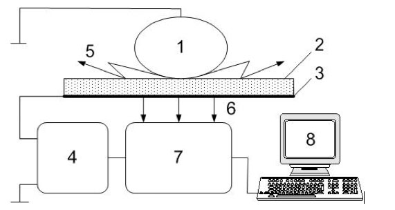
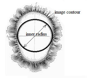
This glow is detected by a sensitive CCD camera (Fig. 1) and processed by the computer as a single digital image. Individual EPI images (Fig. 2) of all ten fingers provide a set of quantitative parameters which may be used for statistical analysis and practical applications [4-15]. Parameters are calculated automatically using image processing of proprietary cloud-based software (www.Bio-Well.com). Through investigation of the fluorescent fingertip images, which dynamically change with emotional and health states, one can identify areas of congestion or health within the whole system. EPI applications are being developed for different areas of research [5-22]. “The data processor represents a specialized software complex, which allows carrying out standardized processing of the fluorescent images: capturing images, filtration of images, obtaining numerical characteristics, creation of graphs and diagrams, saving data and transfer of data for additional processing” [11].

Schematic representation of the Electrophotonic Imaging technology. 1 – object of the study; 2 – glass electrode; 3 – transparent conductive cover; 4 – impulse generator; 5 – gaseous discharge; 6 – photon emission; 7 – optical system; 8 – computer.

EPI image of a finger with some indicated parameters.
Each generated electrophotonic image is analyzed by sector division, according to acupuncture meridian correlations developed by Dr. Peter Mandel in Germany [23]. “The parameters of the image generated from photographing the finger surface under electrical stimulation creates a neurovascular reaction of the skin, influenced by the nervous-humoral status of organs and systems. Due to this, the images captured on the EPI register an ever-changing range of states. In addition, most healthy people’s EPI readings vary only 8-10% over many years of measurements, indicating a high level of precision in this technique” [4].
One of the first studies of cancer diagnosis with EPI technology was the work of B.L. Gurvits et al. [24]. The material for the study were plasma samples of patients with cancer of various organs, both with the absence and the presence of distant metastasis, as compared to blood samples of healthy donors. It was found that for all the samples the values of discharge parameters of blood for cancer patients were significantly higher than values for healthy people. R.S. Chouhan et al. [25], examining the EPI-grams of fingers of patients with different stages of cervical cancer, have shown their significant difference from healthy patients on the images parameters. R. Vepkhvadze et al. [26] by EPI monitoring of patients with squamous cell lung cancer, showed that the results of EPI evaluation and monitoring of the functional status of the patients correlate with clinical, laboratory and instrumental studies in 90-96% of cases. W. Seidov [27] identified some correspondence between EPI parameters and the presence of tumors in different parts of the colon. However, systematic research related to the diagnosis of colon tumors using EPI-graphy has not been found. EPI is a screening procedure and its use for the diagnosis of the colon tumors may help identify patients in the early stages of the disease. This is important because patients often seek help when an oncological disease is already at stage 3-4.
2.2. Subjects Selection
“The Institutional Review Board approval of the consent form was obtained according to the guidelines prescribed by the Review Board. All participants were residents of Moscow. The participants of the study were informed that the results would be published in a journal, or used to teach others. The participants were asked to sign the consent form where it was emphasized that their participation was voluntary and that they could withdraw at any time. Before signing the consent form, the participant could refuse to take part in the study” (https://bioethicsarchive.georgetown.edu/nbac/clinical/Chap3.htm).
137 people were studied with the EPI camera, with ages ranging from 16 years to 86 (56.1 ± 1.5 years); 49 males and 88 females. For all people visual colonoscopy was performed using Evis Exera-II and Exera-III systems (Olympus Co, Japan). Based on the results of the colonoscopy and histological findings all subjects were divided into 2 groups: control group of 55 people (43.3 ± 2.2 years); 9 males, 46 females; and patients with tumors (benign or malignant) of the colon - 82 people (64.2 ± 1.3 years); 40 males and 42 females. Then all subjects were divided into smaller groups based on: morphology (hyperplastic polyps - 23 people, adenoma - 41 people, cancer - 13 people); size (tiny 1-5mm - 21 people, small 5-10 mm - 34 people, medium 10-25 mm - 5 persons); the number of tumors (single - 33 people, multiple - 30 people); localization (right half of the colon - 21 people, left divisions - 34 people, both sides - 9 persons).
2.3. Data Processing
Computer recording and analysis of the EPI images were performed using the "EPI Camera" and “Bio-Well” devices using special EPI and Bio-Well software (www.ktispb.ru, www.bio-well.com). The obtained data were entered into the Excel program and then statistically processed by SPSS Statistics 17.0. The Kolmogorov-Smirnov test, Student t-test, Mann-Whitney U-test, ROC-analysis, discriminant analysis and binary logistic regression were used (http://itl.nist.gov/div898/handbook/index.htm).
The study was conducted using impulse analyzer "Bio-Well" based on Gas Discharge Visualization (GDV) technique. Each participant was asked to place fingers correctly on the glass surface. The pictures of electrophotonic emission of all ten fingertips were taken with a single capturing. The following parameters of the images were found to be the most significant [3]:
Area. Amount of light quanta generated by the subject in computer units - pixels (the number of pixels in the image having brightness above the threshold).
Normalized Area. The ratio of the image area to the area of the inner oval.
Intensity. Averaged intensity of light emission in computer units.
Inner Radius. Radius of the circle inscribed in the inner oval.
Form Coefficient. Calculated according to the formula: FC = k*L2/S, where L is the length of the image external contour and S is the area.
Inner Noise. Amount of pixels in the inner oval [3].
Selection of the most informative parameters for the separation of the control group and patients with colon tumors was performed using the Student's t-test and ROC-analysis. To construct the decision rules stepwise discriminant analysis and binary logistic regression were used.
3. RESULTS
First, the parameters that significantly separated (p < 0.05) groups of healthy individuals and patients were selected with the Student's t-test. As we see from Table 1, different parameters have different trends from group to group. Based on this with the stepwise discriminant analysis decision rule was constructed, which included the following parameters: area, average intensity of the glow, radius of the inscribed circle and the form coefficient. The most significant in the analysis were the sectors of the coccyx, sacrum and lumbar areas (the colon is innervated from these parts of the spine). The specificity of the resulting function was 78.2% and sensitivity 76.8%. Unfortunately, it is impossible to identify the localization of tumors of the colon using this method.
Patterns of change in the parameters of the study groups with increasing degree of tumor neoplasia (average values).
| Parameter | Control | Polypus | Cancer |
|---|---|---|---|
| Control > Polypus > Cancer | |||
| Normalized area | 1.41 ± 0.12 | 1.27 ± 0.06* | 1.09 ± 0.04** |
| Inner noise | 40.90 ± 3.00 | 31.11 ± 2.51* | 23.32 ± 2.01* |
| Isoline radius | 14.21 ± 0.45 | 11.46 ± 0.32* | 10.45 ± 0.42* |
| Intensity | 86.65 ± 0.12 | 78.04 ± 0.08* | 75.19 ± 0.05* |
| Control < Polypus < Cancer | |||
| Inner circle radius | 46.05 ± 1.53 | 54.45 ± 1.63* | 59.37 ± 1.04* |
| Form coefficent | 11.14 ± 0.54 | 17.46 ± 0.60* | 20.52 ± 0.45** |
| Isoline fractality | 1.60 ± 0.02 | 1.63 ± 0.04* | 1.71 ± 0.01* |
| Isoline entropy | 1.57 ± 0.03 | 1.65 ± 0.02* | 1.74 ± 0.01* |
| Isoline length | 950 ± 27 | 1025 ± 16* | 1105 ± 40* |
| Area | 9620 ± 225 | 10760 ± 215* | 11427 ± 115* |
(* p <0.05, ** p <0.001).
As one can see from Table 1, parameters have different tendencies in the line Control => Polypus => Cancer. In order to evaluate the effectiveness of using the method of gas discharge visualization for the detection of neoplasms of the colon, 69 of the most significant parameters for the pathogenesis of the colon tumors were selected using T-student criterion. Stepwise discriminant analysis in the SPSS Statistics 17.0 package was then carried out using data of patients with neoplasms of the colon and the control group.
Included in the result of stepwise discriminant function analysis were the most significant parameters affecting the assignment of patients to one group or another. Namely, the parameters associated with the descending colon, lumbar, sacrum and coccyx.
The equation consisted of 7 variables (Table 2):
I2L - average intensity of the sector of the descending colon of the index finger of the left hand.
I2LCo - average intensity of the coccyx sector of the index finger of the left hand.
S2Lsac - size in pixels of the sacrum sector of the index finger of the left hand.
I2Lsac - average intensity of the sacrum sector of the index finger of the left hand.
R2RLumb - radius of the inscribed circle of the lumbar sector of the index finger of the right hand.
S2Rsac - size in pixels of the sacrum sector of the index finger of the right hand.
F2Rsac - form factor of the sacrum sector of the index finger of the right hand.
The coefficients of the canonical Fisher's linear discriminant functions.
| I2Lav | I2LCo | S2Lsac | I2Lsac | R2RLumb | S2Rsac | F2Rsac | Constant | |
|---|---|---|---|---|---|---|---|---|
| Control | .630 | .598 | -.015 | .501 | 1.108 | .006 | .490 | -94.303 |
| Cancer | .722 | .530 | -.009 | .407 | 1.173 | .009 | .566 | -102.081 |
The classification results obtained by using discriminant analysis.
| Original | Cross-validated | |||||||
|---|---|---|---|---|---|---|---|---|
| Count | %% | Count | %% | |||||
| Specificity | Sensitivity | Specificity | Sensitivity | Specificity | Sensitivity | Specificity | Sensitivity | |
| Control | 47 | 14 | 85.5 | 17.1 | 43 | 19 | 78.2 | 23.2 |
| Cancer | 8 | 68 | 14.5 | 82.9 | 12 | 63 | 21.8 | 76.8 |
| Total | 55 | 82 | 100 | 100 | 55 | 82 | 100 | 100 |
- Cross validation is done only for cases in the analysis. In cross validation, each case is classified by the functions derived from all cases other than that case.
- 83.9% of original grouped cases were classified correctly.
- 77.4% of cross-validated grouped cases were classified correctly.
As we see from the classification matrix of Table 3, the specificity of the resulting function, after cross verification, is 78.2%, and the sensitivity is 76.8 %. We can conclude from these data that the separation between sick and healthy individuals has a fairly high level of precision for screening studies.
We decided, for comparison, to determine the sensitivity and specificity of the method using logistic regression. To achieve this goal the same 69 most significant parameters for the pathogenesis of tumors of the colon that were previously selected using T-student criterion were taken. The values of sensitivity and specificity were calculated at Step 6 as 76.4% and 85.0% respectively (see Table 4).
The classification results obtained using binary logistic regression.
| Control/Patients | all %% | Control/Patients | all %% | Control/Patients | |||||
|---|---|---|---|---|---|---|---|---|---|
| Specificity | Sensitivity | Specificity | Sensitivity | Specificity | Sensitivity | ||||
| Step 1 | Step 2 | Step 3 | |||||||
| Control | 33 | 15 | 33 | 14 | 36 | 16 | |||
| Cancer | 22 | 65 | 22 | 66 | 19 | 64 | |||
| Correct %% | 60.0 | 81.3 | 72.6 | 60.0 | 82.5 | 73.3 | 65.5 | 80.0 | 74.1 |
| Step 4 | Step 5 | Step 6 | |||||||
| Control | 38 | 10 | 39 | 14 | 42 | 12 | |||
| Cancer | 17 | 70 | 16 | 66 | 13 | 68 | |||
| Correct %% | 69.1 | 87.5 | 80.0 | 70.9 | 82.5 | 77.8 | 76.4 | 85.0 | 81.5 |
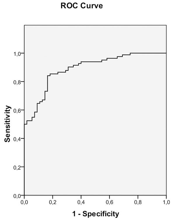
Based on logistic regression ROC-curve was built (Fig. 3).
4. DISCUSSION
We compared the results obtained with the results of other screening methods used for diagnosis of colon tumors. The most well-known screening test - FOBT – is a definition of a small amount of occult blood in the intestinal contents. It is performed at home for 3 days, during which it is necessary to follow a diet without animal protein. Another method of immunochemical test for fecal occult blood, FIT, is more convenient and does not require a special diet for its production as fewer stool samples are needed. According to Kronborg O., et al. [28], “the key for early detection of polyps and colon cancer is an organization of screening of people older than 50 years for occult blood in the stool and endoscopic examination of persons with a positive result of this analysis; this approach ultimately reduces mortality from colorectal cancer by 15-33%”. However, this method cannot be used in the presence of hemorrhoids. According to [29] “the sensitivity of various noninvasive diagnostic methods for detection of the colon polyps ranges from 30 to 95%” (Fig. 3). However, these methods miss the small non-bleeding polyps. Often they produce false-positive and false-negative results. Colonoscopy screening is referred to as the gold standard in some countries. Colonoscopy allows inspecting the whole colon and removing detected polyps. However, “the method is time consuming, rather expensive, requires bowel preparation, it is unpleasant for the patients” [30]. Virtual colonoscopy avoids painful bowel preparation performed in conventional colonoscopy. The sensitivity of this method in the diagnosis of polyps larger than 10 mm is 90%, while 80% for polyps of 5-9 mm in size, and 67% when the size of the polyp does not exceed 5 mm. Specificity of the method depends on the size of the tumors [2]. However, along with advantages, the method of virtual colonoscopy has significant shortcomings, such as financial inaccessibility and the inability to perform a biopsy, thereby yielding a standard colonoscopy. With EPI technology we obtained sensitivity from 74% to 85%, and specificity from 66% to 77% (Fig. 4). Thus, the results have proven the ability to identify patients with tumors of the colon using EPI technology, as well as use it for differential diagnosis of tumors of the colon by their morphology, size and quantity. Of course, this is only a preliminary study and much research is needed to find a reliable method for detecting colon tumors using the EPI technique. We need to point out that EPI testing in non-invasive, takes less than five minutes, and the equipment is relatively cheap and accessible. This opens up good prospects for further research for Electrophotonic Imaging analysis implementation as a first step of the screening process.

ROC curve. Parameters: Area under the ROC-curve is 0.891; Standard deviation error under the nonparametric assumption 0.027; lower and upper 95% confidence intervals 0.839 and 0.944, respectively.
When comparing the results of decision rules obtained by using discriminant analysis and binary logistic regression, it should be noted that the specificity of the second rule is somewhat lower (78.2% compared to 76.4%), but the sensitivity is much higher (76.8% and 85.0%). This suggests that although the first is a little better at revealing healthy people, the possibility of identifying the really sick people among the surveyed is significantly higher using logistic regression.

Characteristics of different methods of non-invasive diagnosis of colon polyps [29].
It should be noted that among the 7 factors included in the discriminant function, and 6 included in the logistic regression equation, 5 are the same, which confirms the high quality of the obtained functions.
This paper presents the pilot study developing methodological approach to the GDV data processing. That is why we tried different methods of data processing. At the same time we do not pretend to develop a diagnostic method – sample size is too small for this, and other cancer types were not studied. Further research is needed.
CONFLICT OF INTEREST
The authors confirm that this article content has no conflict of interest.
ACKNOWLEDGEMENTS
Declared none.


